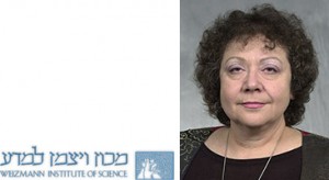
Фото: профессор Лея Айзенбах
www.annashats.com
Иммунитет против рака — в Израиле начаты испытания вакцины нового поколения. Записки Фиговского.
На днях в онкологическом центре «Давидов» при больнице «Бейлинсон» в Петах-Тикве начались клинические испытания нового препарата. По словам руководителя экспериментальной группы проф. Нира Пеледа, первые результаты превзошли все ожидания. Если так пойдет и дальше, то уже в 2015 году израильские ученые смогут заявить, что они сумели окончательно победить рак.
Открытие израильских ученых доказывает, что случаи чудесного исцеления от рака без лекарств отнюдь не следует огульно относить к шарлатанству, медицинским ошибкам и т.п. Дело в том, что наша иммунная система действительно вполне способна бороться с раковыми клетками и уничтожать их. Правда, эта способность в немалой степени связана с индивидуальными свойствами организма и уменьшается с возрастом. Но если бы наш организм не имел возможности уничтожать раковые клетки, проникающие в него тем или иным путем, все человечество поголовно умирало бы от рака.
Объяснять весь биохимический механизм действия новых белков непосвященному человеку трудно, но проф. Айзенбах пользуется для этого прекрасным бытовым примером. Представьте себе, говорит она, что в некий город прибыла банда известных грабителей. Все документы у них в порядке, они отлично маскируются под обычных законопослушных граждан, так что у полиции нет никакого формального повода для их ареста и отправки за решетку. Больше того, полицейские не могут распознать этих бандитов в толпе. Но тут в городе появляются частные детективы, которые проводят собственное расследование, устанавливают личности бандитов и передают сведения о них полиции. Теперь полицейские могут начать действовать.
Созданные израильскими учеными белки выступают в роли тех самых частных детективов, которые указывают защитной системе нашего организма на проникших в нее убийц, после чего та успешно ликвидирует их собственными силами. Как отмечается, новые препараты уже прошли целый ряд успешных проверок, в том числе и на людях.
На самом деле значение открытия израильских ученых куда шире, чем представляется на первый взгляд. Ведь если самое главное заключается в том чтобы помочь нашей иммунной системе опознавать раковые клетки, это означает, что белки-“детективы» можно ввести в организм заблаговременно, и тогда они помогут распознать раковые клетки на самой ранней стадии их появления в организме. Проще говоря, это открытие позволяет реализовать самую заветную мечту медиков — создание прививки от рака. Правда, следует помнить, что формально называть это прививкой нельзя, так как у нее иной, прямо противоположный механизм действия, чем у других видов прививок, заключающихся в ведении в организм ослабленных вирусов того или иного заболевания.
К тому же проф. Айзенбах не исключает, что такая «прививка» будет обладать временным ограничением действия, и время от времени ее придется повторять.
Проф. Олег Фиговский, Технион, Израиль
На сайте профессора сообщается, что работа ведется пока в основном с животными, так как идет стадия научной разработки. Однако главный принцип — дополнительного обучения имунных клеток уже понятен и по всей видимости стадия практического медицинского применения в клинике Davidoff — центра в Израиле уже наступила.
Подробнее — научное сообщение профессора Леа Айзенбах:
Cancer, Metastasis and Immunotherapy: Identification of tumor associated antigens, vaccination strategies, mechanisms of anti-tumor immunity, tumor escape and tolerance, cryo immunotherapy, the role of small interferon induced genes in tumorigenicity and apoptosis, TCR evolution.
The treatment of tumors, especially of metastasis by chemotherapy or irradiation often fails. A variety of biological treatments aimed at inhibition of cell growth or angiogenesis, or at induction of apoptosis are being developed. Additional approaches seek to activate the immune system to destroy tumor cells.
One of these approaches, aimed at activation of T cells, is based on the notion that qualitative and quantitative differences in the antigenic repertoire between tumor cells and normal cells can be recognized by the T cell repertoire that contains anti-self clones and can be efficiently activated. This antigenic repertoire also named Tumor Associated Antigens (TAA) consists of mutated proteins, embryonal re-expressed proteins, normal overexpressed proteins, tissue specific proteins or in rare cases of virally induced tumors, of viral proteins. The peptides derived from processing of these proteins are presented by MHC class I to Cytotoxic T Lymphocytes (CTL) or by MHC class II to T helper cells (TH cells).
Using reverse immunology and a screening system based on Db-/-Xbeta2 Microglobulin double knockout mice, transgenic for a single-chain HLA-A2/Db-beta2 Microglobulin molecule (HHD mice), we have identified novel immunogenic and antigenic TAAs from prostate, breast, colon and bladder carcinomas. Specific CTL were derived by immunization of HHD mice with tumor peptide extracts or with synthetic nonapeptides loaded on antigen presenting cells or with HHD transfected human tumor cell lines. These CTL reacted with series of peptides derived from overexpressed candidate proteins, identified by differential display methods (SAGE, Microarrays, SSH). Comparison of CTL derived from HHD mice to CTL induced from patient’s PBMC showed overlapping recognition of many candidate peptides. Peptide vaccines, derived from the prostate antigens PAP and STEAP induced CTL that retarded growth of human tumors xenografts in adoptive transfer experiments. A novel HHD tumor model for carcinomas was used in active immunization experiments. Preventive immunization and therapeutic vaccination prolonged mouse survival. Similarly, CTL to peptides derived from lactadherin (BA46-1) retarded xenografts of breast carcinoma. Peptides and Glycopeptides derived from MUC1, highly overexpressed in breast carcinomas, showed overlapping activities in HHD mice. Yet, in HHDXMUC1 compound mice, only one highly immunogenic peptide (D6) retained the ability to activate effectively CTL. Novel Bladder Carcinoma (TCC) TAA peptides were defined from the overexpressed proteins MAGE-A8 and Cripto-1 and from transitional cell tissue specific proteins, the uroplakins.
Analysis of colon carcinoma differentially expressed genes by SAGE, followed by screening for HLA-A2 binding peptides resulted in 500 candidate peptides for immunogenicity screening. We have identified 22 antigenic peptides of which 7 peptides were found to be immunogenic in HHD mice and human PBMC. Interestingly 3 of these peptides are derived from the same protein, the 1-8D interferon (IFN) inducible protein, which belongs to the 1-8 interferon-inducible gene family. Three members of the family 1-8D, 1-8U and 9-27 are linked on an 18Kb fragment of chromosome 11 and are highly homologous. The relative expression of the human 1-8D gene in normal and tumor colon tissues, derived from colon cancer patients, using real time PCR, demonstrated that the 1-8D gene is overexpressed 4-16 fold, in 9/13 tumor samples compared to paired normal tissue. Sequence comparison of the human 1- 8D in pairs of tumor/normal colon samples revealed 6 different alleles containing single nucleotides polymorphisms (including insertion) in the 1-8D gene. We have constructed variant human 1-8D cDNAs fused to HA tag or to GFP protein for further analysis. Cell cycle analysis of NIH 3T3, CT26 colon carcinoma, HeLa HCT116/TP53+/+, HCT116/TP53-/- and H1299 cells transfected with 1-8D showed elevation in the G1 fraction of the cells, 24hr following transfection, and elevation in the sub G1 phase of the transfected cells 48 and 72hr following transfection. Annexin V binding assays and TUNEL assays showed increased populations of apoptotic cells in cells transfected with 1-8D compared to cells transfected with empty vector or without DNA, 48hr following transfection.
Apoptosis is mediated by caspase 3 and 9 intrinsic or mitochondrial mediated pathway. Althought 1-8D induces apoptosis independent of TP53, TP53 expression downregulates 1-8D expression on the protein level. Transformation assays revealed a possible role of 1-8D gene as a transformation inhibitor. Another interferon inducible gene, the ISG12 was found to be highly overexpressed in a series of immortalized breast carcinoma lines compared to immortalized normal breast epithelium lines by SSH and by microarray analyses. ISG12 is a member of a family of small intracellular non-secreted proteins (10-20 kDa), and has high homology to the 6-16 protein, to which there are indications of a direct role in growth inhibition. Like the 1-8D, we found that ISG12, overexpressed in 60% of breast carcinoma lines, shows proapoptotic effects in transiently transfected cells. The significance of overexpression of proapoptotic genes in tumors is under investigation. Immunotherapy, either by active vaccination or by adoptive transfer of immune cells, often reduces the growth rate of established tumors and prolongs survival, yet complete elimination of tumors is rare. A number of hypotheses were suggested to explain this failure: A. The speed of tumor proliferation versus a limited number of specific T cells. B. Immune editing-creation of antigen loss variants or MHC/TAP loss variants. C. Inactivation of immunocytes by products of the tumor. D. Failure of immunocytes to reach the tumor.
To investigate the dominant factors that reduce anti-tumor immunity we used the most extreme model available: The OTI transgenic mouse has a CTL population which is essentially monoclonally specific for the high affinity MHC class I-Kb binding, Ovalbumin (Ova) derived peptide SIINFEKL. The B16- MO5 melanoma is stably transfected with the Ova gene and is therefore efficiently recognized and lysed by OT-I CTL. A further advantage of this system is the xenograft nature of the Ova antigen, which is not tolerized against. By using the OT-I/MO5 model we show that even when ~100% of T cells recognize the tumor it continues to grow. We also show that the tumor utilizes an «antigen loss» escape mechanism by downregulating expression of the Ova gene. We further demonstratethe importance of a polyclonal CD8 T cell repertoire in tumor rejection. Yet, the tumor itself has a suppressive effect on T cells.
In collaboration with Prof. Irun Cohen, we investigated the possibility of inducing autoimmunity to tissue specific antigens as a mean to attack tumors of the same tissue, using melanoma, glioma and astrocytoma models. Breaking tolerance was shown to be possible by lymphoabalative treatments followed by active immunization with tissue specific proteins or their derived peptides or by passive transfer of CD4 T cell lines specific to tissue restricted proteins. In another project we have shown a phenomenon we term ‘split immunity’ in which a live tumor challenge in one site (especially an immune privileged site) is lethal, while it’s placement in another site, generates immunity. This immunity could spread to the original site in which the tumor is lethal, increasing animal survival and in some cases causing complete tumor regression. We have shown this phenomenon both in protection assays, immunizing the animals before challenge, and in immunotherapy tumor rejection assays. While immunotherapy through active defined peptide vaccines, T cell transfer or manipulation of tolerance are single modality approaches a combined approach of cryosurgery and immunotherapy was tested in metastatic tumor models. In situ administration of immature Dendritic Cells (DC), following cryotherapy of primary tumors was shown to induce systemic CTL and TH1 responses and eradicate metastasis.
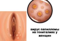
Иллюстрация: www.gazeta.ru
http://www.ecolife.ru/zdorove/40041/


